Glaucoma – a deep dive into the condition
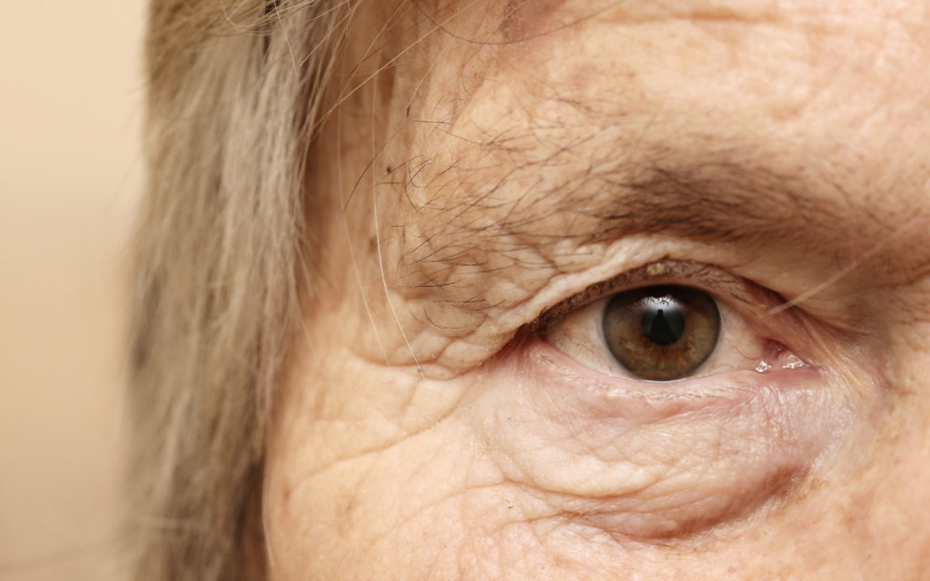
2% of the population over the age of 40 have glaucoma and worryingly half of these people don’t even know they have it. This disease is most prevalent in the elderly community, however people who are short sighted, diabetic or have a family history of glaucoma are also at a higher risk. Glaucoma will often affect both eyes, however one eye may develop the disease quicker than the other. If it is left untreated it can lead to blindness.
Mr Vik Sharma, consultant ophthalmic surgeon at the HSEH says “Glaucoma is a disease of the optic nerve”. More specifically, glaucoma is a group of conditions. Most types of glaucoma are associated with increased pressure within the eye, which in turn puts pressure on the optic nerve, damaging your sight. However it can occur when the pressure is not higher than normal, but the nerve damage still occurs and the resulting loss of vision.
What are the different types of glaucoma?
One of many types of glaucoma may affect an individual. The different types are:
- Primary open-angle glaucoma – This is the most common and most chronic type of glaucoma. The aqueous humour is a thin, transparent fluid similar to plasma that flows out of the eye through web-like channels. These channels ensure that excessive pressure isn’t built on the optic nerve. The primary open-angle glaucoma occurs when the drainage canals in the eye do not work as they should and the pressure within the eye and on the optic nerve increases.
- Primary angle-closure glaucoma – In this condition, the drainage canals in the eye are blocked by the iris and the pressure on the optic nerve is constantly high. The iris comes forward, away from its place, and closes-off the drainage canal.
- Secondary glaucoma – When a different eye condition causes injury to the optic nerve or leads to an increase in pressure on the optic nerve, it results in secondary glaucoma. Here, glaucoma is a consequence of another serious eye condition.
- Congenital glaucoma – This glaucoma condition occurs in babies and children. It could either be hereditary or due to an eye condition or injury that occurs due to foetal abnormalities during the pregnancy.
- Iridocorneal endothelial syndrome – This is a very rare type of glaucoma where cells on the posterior surface of the cornea disintegrate and bind like adhesions to the iris. This can cause the surface of the iris to get damaged while the drainage canals in the eyes become clogged.
- Other conditions – Apart from the conditions above, glaucoma can also occur if a chemical is sprayed into the eye and burns the optic nerve. Additionally, any bacterial, viral or fungal infection in the eye may sometimes damage the optic nerve, causing glaucoma.
Around half of those that are suffering with glaucoma are unaware that they have it, because it can develop without presenting any serious symptoms; until there is a serious change in vision. One of the hardest parts of treating glaucoma is finding it early enough so that it does not cause any visual loss. As a country we should encourage people to have regular eye tests so that the disease can be detected more easily and earlier. People should aim to have an eye test once every two years as a bare minimum.
At the moment the standard test for glaucoma include an air puff test which tests the pressure in the eye. This test can miss up to 60% of cases as not all patients will present with high pressure in the eye. Even if the disease does cause an increased pressure, it may not be caught by the test as the pressure in your eye can fluctuate.
If you know that you have a family history of glaucoma then it is important that you bring this to the attention of your optician so that they can look at in in more detail. As glaucoma often causes loss of peripheral vision, they may perform a test to look at your field of vision.
The damage that is caused by glaucoma cannot be reversed so it is important that the disease is caught early and managed properly. For patients that are treated at an early stage it is unlikely that they will ever go on to develop any damage to their vision.

Diagnosis
At HSEH we conduct a series of specialist examinations and tests when checking for glaucoma. These can include:
- Nerve scans – the scan is used to look at a specific layer of the nerve, this can help detect early damage
- Visual field test- uses the latest short-wave analysis perimetry software to detect the disease up to 5 years earlier than previously possible
- Ocular blood flow measurement – glaucoma can lead to a reduced flow of blood, and therefore oxygen, to the optic nerve so monitoring this flow can give a good indication of glaucoma
- Eye pressure – using an applanation tonometry machine you can get a more precise result than from a standard
- Corneal pachymetry ultrasound – this measures the thickness of the cornea to determine how accurate the pressure readings are as the thickness of the cornea has been shown to influence eye pressure readings.
So how do we treat glaucoma?
The key to successful treatment of glaucoma is usually associated with lowering pressure within the eye and therefore reducing the risk of permanent damage. There are a number of options when it comes to treatment.
- Eye drops – these eye drops contain a chemical which is already found in the body, that effectively lowers pressure in the eye in less severe cases. This is not only safe, but also convenient and easy for most of our patients.
- Laser treatment – helps to facilitate the outflow of fluid from the eye through the normal channels. The laser that is used at HSEH is extremely safe and painless. Around 80% of patients have effective results from this procedure. As the effects of this treatment are not permanent and usually only last a few years, you will have to attend regular appointments to monitor your eye pressure, at least every 6 months.
- Surgery – although the risks associated with surgery are very low this will usually only be offered as a last resort if other treatments have failed. This is considered the gold standard for treating the condition. During the surgery a channel is made to allow the fluid to drain through the wall of the eye into the skin of the eye. The body will, naturally, try to heal the new channel, however we will provide anti-scarring agents to ensure that this doesn’t happen. There are also micro-implants which maintain the channel and enhance the recovery process.
Read more about your eye care at HSEH here.

0 Comments